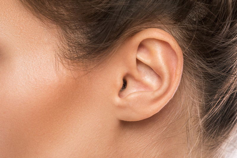
The inner ear is a remarkable sensory organ responsible for our ability to hear and maintain balance. It is composed of a complex network of cells and structures that work together to detect and interpret sound waves and movement.
Recent research has uncovered the fascinating phenomenon of cell self-organization in the inner ear, whereby cells are able to organize themselves into precise patterns and structures without external guidance or intervention. This self-organization is essential for the development and function of the inner ear, and understanding the mechanisms involved could have significant implications for the treatment of hearing and balance disorders.
The inner hair cells are the primary sensory cells within the Corti organ, responsible for detecting sound vibrations and converting them into electrical signals. The outer hair cells, on the other hand, play a role in amplifying and sharpening the sound signals detected by the inner hair cells. The supporting cells provide structural support and metabolic functions to the hair cells.
The Corti organ is organized into several layers, with the inner hair cells located closest to the center of the cochlea and the outer hair cells located further out. The precise arrangement of these cells is essential for the detection and interpretation of different frequencies of sound.
Overall, the Corti organ is a remarkable structure that plays a critical role in our ability to hear and interpret sound. Its complex organization and specialized functions highlight the sophistication and precision of the mechanisms involved in auditory perception.
The checkerboard arrangement in the inner ear refers to the pattern of organization of the sensory cells responsible for detecting sound waves and translating them into electrical signals that the brain can interpret. Specifically, in the cochlea of the inner ear, the sensory cells responsible for detecting different frequencies of sound are arranged in a pattern that resembles a checkerboard.
The hair cells are arranged in rows along the length of the cochlea, with different rows responding to different frequencies of sound. Within each row, the hair cells are arranged in a checkerboard pattern, with adjacent hair cells responding to slightly different frequencies of sound. This arrangement allows for precise discrimination of different frequencies of sound, which is essential for hearing and speech perception.
The checkerboard pattern in the inner ear is an example of the remarkable precision and organization of biological systems, and it highlights the complexity and sophistication of the mechanisms involved in our ability to hear and interpret sound.
A recent study conducted by a Japanese research group revealed the checkerboard pattern?s crucial role in hearing. In studying mice, the researchers found that abnormalities in the inner ear resulted in hearing loss. This indicates that further studying the patterns of the inner ear and cell self-organization could lead to a greater understanding of hearing loss and its causes. Furthermore, this checkerboard pattern is found in other sensory organs, including the olfactory epithelium that is responsible for the sense of smell and the retina which is responsible for vision. Additional research could help us better understand sensory organs, how they function, and the diseases and abnormalities that affect them.
For more information about this intriguing new research, we invite you to contact our hearing practice today.


